Hip replacement is a surgery recommended for people with severe hip damage. When a hip replacement is done, the surgeon removes damaged cartilage and bone from your hip joint and replaces them with new, man-made parts. This helps your hip joint function better, relieves pain and improves your walking and other movements. This is usually advised when physical therapy and medications have failed to improve the condition.
Osteoarthritis and Hip Replacement
Osteoarthritis of the hip is the most common reason for a hip replacement. Aging causes wear and tear of the cartilage covering the joint surfaces resulting in great pain and stiffness. This condition is called as Osteoarthritis.
Destruction of the hip joint can also be caused due to loss of the blood supply to the head of the thighbone (osteonecrosis), rheumatoid arthritis, injury, infection and developmental abnormalities of the hip. Patients with arthritis may also have brittle bones (osteoporosis), but there is no direct relationship between bone density and the development of arthritis of the hip.
Symptoms
Hip arthritis typically causes pain that is dull and aching. The pain may be constant or it may come and go. Pain may be felt in the groin, thigh and buttock with possible referred pain to the knee. walking long distances can also cause severe pain resulting in the patient limping. Some patients may need a cane, crutch, or walker to help them get around. Pain usually starts slowly and worsens with time and higher activity levels.
Patients with a bad hip joint can have difficulty in certain normal ativities like climbing stairs, wearing trousers, tying shoes and clipping toenails.
Diagnosis
X-rays may show loss of the cartilage space in the hip socket and a "bone-on-bone" appearance along with bone spurs and bone cysts. A magnetic resonance imaging (MRI) or computed tomography (CT) scans may be recommend to confirm the diagnosis.
Nonsurgical Treatment
For hip arthritis, the first treatment a doctor may recommend is over-the-counter, anti-inflammatory medications. Certain nutritional supplements including glucosamine, could provide some relief. Short-term physical therapy may help improve strength and reduce pain. Transferring of weight to a walker helps relieve pain and walking ability.
Surgical Treatment
General anesthesia is most used for joint replacement surgeries but sometimes regional anesthesia is also used. This depends on your doctor, on your overall health and personal preference.
The damaged cartilage and bone is first removed. To remove the worn out ball of the ball-and-socket hip joint, the bone is cut to remove the femoral head. In order to insert a new joint, the damaged bone and cartilage must first be removed. Once the arthritic ball is removed, the worn out socket can be addressed. Unlike the ball, this bone cannot be cut off -- the socket of the hip joint is part of the pelvis bone.
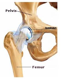
A reamer is used to scrape away the damaged cartilage and bone and a smooth, perfectly rounded surface is got which accepts the new hip implant. Once the damaged bone has been removed, the new socket of the hip replacement can be inserted. The socket of the pelvis is called the acetabulum and the part of the hip replacement inserted into the socket is called the acetabular component. The acetabular component is held tightly in the pelvis by making the socket slightly smaller than the acetabular component and wedging the implant into the bone. The implant has a rough surface to allow bone to grow into the surface of the implant over time.
Now that the socket has been addressed, attention can turn to the ball of the ball-and-socket hip joint. The ball is supported with an implant inserted down the hollow center of the thigh bone (femur). This implant is called the femoral stem.
For the femoral stem to be held tightly in the bone special tools are used to shape the center of the thigh bone to accommodate the femoral stem. With the bone prepared, the femoral stem is inserted and held in the bone with or without cement.
If bone cement is used, the cement is inserted in a liquid form and the stem is then placed. The cement permanently hardens within a few minutes to hold the implant fixed within the bone. When no cement is used, the implant is called "press-fit." This means that the implant is wedged tightly into the bone. A rough surface covering the implant allows bone to grow into the implant over time.
With the stem inserted down the center of the thigh bone, the ball of the ball-and-socket hip joint can be inserted on top of the stem. A metal ball is tightly fit onto the top of the stem.
Pain medication will be given depending on the severity of the pain. Anticoagulants may be prescribed for several weeks following surgery.
On a cemented joint, mobilization can be done and partial weight loading is done within 24 hrs. On an un-cemented hip for weight bearing is not permitted for about 6 weeks. With a cemented or hybrid (one piece cemented and one piece un-cemented) hip, you can usually put some weight on your leg right away, but you'll still need a walker, a cane, or crutches for several weeks.
Precautions
The other Orthopedic Procedures are:
Few Major Hospitals for Hip Replacement are:
Thailand, Malaysia, Singapore, Turkey and India are the most cost effective locations that offer up to almost 80% savings in comparison to the US.
SurgeryPlanet facilitates a plethora of services to the medical treatment traveler also which includes, a hassle free and discounted travel option, a welcome hand at the airport on arrival, travel in an air-conditioned car, round the clock service & support. Your medical evaluation is pre arranged with the least of waiting time. Once your assessment is complete and found medically fit, the procedure is immediately scheduled without a waiting period. Please read through our Services and Testimonials to understand and select your best options.
Major Treatments Abroad: Obesity / Bariatric Surgery | Spine Surgery | Stem Cell therapy | Fertility treatment | Knee replacement in India and Thailand | Heart Surgery | Organ transplant | Ayurveda Treatment | Heart valve replacement | Hip resurfacing | Hospitals in India and Thailand for Laparoscopic Sterilization| Best hospitals in Asia | JCI & ISO certified Hospitals | Cost effective medical procedures | Healthcare tourism | Complete privacy for affordable cost | Weight loss procedures | Infertility treatment | Board certified physicians | Low cost surgeries
SurgeryPlanet is an Healthcare Facilitator and not a Medical service provider. The information provided in this website is not to be used for diagnosis or treatment of any medical condition or use for any medical purposes. We provide information solely for medical travel facilitation and do not endorse any particular health care provider, hospital, facility, destination or any healthcare service or treatment listed. We are not an agent for, or affiliated to any health care provider, or service listed in our website and is not responsible for health care services provided by them. Choice of hospital or doctor for your healthcare services is your independent decision. Consult your domestic licensed health care provider before seeking the services of any health care provider you learn about from our website.