Craniotomy is a surgical procedure in which a portion of the skull, or cranium, is removed to access the brain. The surgery is performed to treat various neurological conditions, including brain tumors, trauma (such as skull fractures or brain bleeds), vascular abnormalities, and some cases of infection or epilepsy. Once the issue is treated, the bone flap is generally replaced and secured in place.
A craniotomy is different from a craniectomy, in which the bone is permanently removed and not replaced. Craniotomy allows surgeons to directly address problems within the brain while minimizing potential damage to nearby structures. It is a highly specialized procedure that requires careful planning and execution, and is performed by neurosurgeons who have expertise in brain surgery.
Craniotomy is used for conditions that involve deep or difficult-to-reach areas of the brain, and it offers the potential for significant improvements in health outcomes, depending on the underlying issue. However, because it involves opening the skull, there are risks associated with the procedure, and it requires a careful balance between surgical intervention and preserving brain function.
Craniotomy is often necessary when a neurological issue is present that requires direct access to the brain. The causes that may necessitate a craniotomy are typically related to conditions that affect brain structure, function, or blood flow. Here are some of the primary causes and risk factors associated with craniotomy:
Brain tumors are one of the most common reasons for performing a craniotomy. Tumors can be benign or malignant and may either grow in or around the brain, exerting pressure on vital structures. The craniotomy procedure may be used to remove, biopsy, or debulk the tumor to prevent further damage or to relieve pressure in the brain.
Traumatic brain injuries caused by accidents, falls, or other blunt force trauma often lead to brain hemorrhage, bruising, or swelling. A craniotomy may be required to remove blood clots (hematomas), repair skull fractures, or relieve intracranial pressure caused by swelling.
A brain aneurysm is a weakened area in the blood vessel walls in the brain that can burst, leading to a hemorrhagic stroke. A craniotomy is often performed to clip or seal the aneurysm and stop the bleeding, reducing the risk of further damage.
Brain infections, such as brain abscesses, may require surgery for drainage if antibiotics are insufficient. A craniotomy is used to access the site of the infection and remove infected tissue, allowing for better resolution of the condition.
In some cases, craniotomy is performed as a treatment for severe, drug-resistant epilepsy. In such cases, the surgeon may remove or alter the part of the brain responsible for the seizures.
In patients with hydrocephalus (excess fluid accumulation in the brain), a craniotomy may be performed to create an opening or to place a shunt to drain the fluid and relieve pressure on the brain.
Arteriovenous malformations (AVMs) or other blood vessel abnormalities may require craniotomy to remove or treat the malformation and prevent further bleeding or neurological damage.
Patients who may require a craniotomy often present with a range of neurological symptoms based on the condition that has led to the need for surgery. Symptoms vary depending on the location of the problem in the brain, its severity, and whether it is acute or chronic.
Severe headaches that do not respond to normal pain relief measures can be a sign of increased intracranial pressure due to tumors or bleeding in the brain.
Sudden loss of vision or visual disturbances, such as blurred or double vision, could indicate pressure on the optic nerve or brain abnormalities.
Numbness or weakness on one side of the body, or difficulty moving a limb, could be a sign of a stroke or tumor affecting motor areas of the brain.
Seizures may occur due to a range of causes, including brain tumors, vascular malformations, or traumatic injury. Seizures are a significant indication for a craniotomy when medication is ineffective.
Memory loss, confusion, or significant changes in personality and mood may occur, especially if a brain tumor or injury affects areas of the brain involved in cognition or emotions.
Damage to the areas of the brain responsible for language (e.g., Broca's area) can lead to difficulty speaking or understanding speech. This can occur as a result of stroke, brain tumor, or brain injury.
Dizziness, difficulty walking, or lack of coordination may occur when the cerebellum or other parts of the brain responsible for motor control are affected.
Symptoms like nausea, vomiting, vision changes, and lethargy may suggest increased intracranial pressure from a mass effect (such as a tumor or hematoma).
The diagnosis leading to the need for craniotomy begins with a thorough evaluation of the patient's medical history and a neurological exam. Various imaging tests are used to determine the exact cause of the symptoms and to guide surgical planning.
The doctor will review the patient’s medical history and conduct a thorough neurological examination to assess cognitive function, reflexes, strength, sensation, and coordination.
CT Scan (Computed Tomography): A CT scan is often the first imaging test to quickly assess the brain for any bleeding, swelling, or tumors. It provides detailed images of the brain and is useful in emergencies.
MRI (Magnetic Resonance Imaging): An MRI provides more detailed images of brain tissue and can help locate tumors, brain injuries, or other abnormalities that may require surgical intervention.
Cerebral Angiography: This imaging technique involves injecting a contrast dye into the blood vessels to provide detailed X-ray images of the brain’s blood supply. It is used to detect aneurysms, AVMs, and other vascular abnormalities.
If a brain tumor or abnormal growth is suspected, a brain biopsy may be performed either during surgery or using a needle to obtain tissue samples for analysis.
Craniotomy is performed when conservative treatments are insufficient, and surgery is necessary to address a condition affecting the brain. The specific treatment options depend on the underlying cause of the condition.
For patients with brain tumors, the goal of craniotomy is to remove the tumor or as much of it as possible to alleviate pressure on the brain. This may involve complete or partial resection of the tumor, depending on its size, location, and type.
In cases of traumatic brain injury, a craniotomy may be performed to remove blood clots (hematomas) and relieve pressure on the brain. This helps prevent further neurological damage.
For brain aneurysms, a craniotomy is used to expose the aneurysm and place a clip around the base of the aneurysm to stop blood flow, preventing rupture and reducing the risk of hemorrhage.
In patients with hydrocephalus (excess cerebrospinal fluid in the brain), a shunt may be inserted during craniotomy to drain the fluid from the brain's ventricles to another part of the body where it can be absorbed.
Craniotomy is sometimes used to treat drug-resistant epilepsy by removing or altering the part of the brain responsible for causing seizures.
While craniotomy is often performed as a life-saving procedure, it is essential to prevent conditions that could require such surgery in the first place and to manage the patient’s condition after surgery:
Individuals at high risk of stroke or brain tumors should undergo regular neurological evaluations and imaging tests to detect any changes in the brain early.
Controlling blood pressure and managing high cholesterol can reduce the risk of stroke and brain aneurysms, thereby lowering the chances of requiring craniotomy.
Maintaining a healthy lifestyle through balanced nutrition, regular exercise, and smoking cessation can reduce the risk of stroke, heart disease, and brain-related conditions that may necessitate surgery.
After a craniotomy, pain management, wound care, and post-operative monitoring are essential. The surgical site must be monitored for signs of infection, and neurological functions must be assessed regularly.
While craniotomy is a highly effective treatment, it carries certain risks and potential complications:
Infection at the surgical site is a risk after any surgery. It can lead to further complications and may require additional treatment or antibiotic therapy.
There is a risk of bleeding during or after the surgery. In some cases, a reoperation may be required to manage excessive blood loss.
Nerve damage to areas such as the facial nerve or vagus nerve may occur during surgery, leading to temporary or permanent weakness, numbness, or difficulty speaking and swallowing.
Some patients experience memory loss, cognitive dysfunction, or mood changes following craniotomy, particularly if large areas of the brain are affected by the condition.
Living with the condition that requires craniotomy—such as a brain tumor, aneurysm, or traumatic injury—can be challenging. After surgery, patients may face physical, cognitive, and emotional challenges.
Post-operative rehabilitation is critical for restoring strength, coordination, and cognitive function. Physical therapy, speech therapy, and occupational therapy can help patients regain independence and improve quality of life.
Surgery and recovery can be emotionally taxing, and patients may experience anxiety, depression, or stress. Psychological counseling or support groups can provide much-needed emotional support.
Patients who have undergone craniotomy require long-term monitoring to assess the success of the surgery, manage any complications, and prevent recurrence of the condition that led to the surgery.
A craniotomy is a surgical procedure in which a part of the skull is removed to allow access to the brain. This surgery is typically performed to treat conditions such as brain tumors, brain hemorrhages, aneurysms, infections, or other brain-related disorders. After the surgery, the portion of the skull that was removed is usually replaced and secured with plates or screws.
A craniotomy is needed for a variety of medical reasons, including:
Brain tumors: To remove tumors or biopsies.
Brain aneurysms: To repair or clip an aneurysm to prevent rupture.
Traumatic brain injuries: To repair damage caused by accidents or falls, such as blood clots or swelling.
Infections: To treat brain infections such as abscesses.
Epilepsy: In some cases, craniotomy is performed to remove the part of the brain causing seizures.
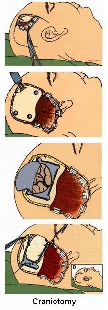
A craniotomy involves the following steps:
Anesthesia: The patient is placed under general anesthesia to ensure they are unconscious and pain-free.
Incision: The surgeon makes an incision in the scalp, and a portion of the skull (bone flap) is removed to access the brain.
Surgical intervention: Depending on the condition being treated, the surgeon will perform the necessary procedure, such as removing a tumor, repairing a blood vessel, or draining a fluid buildup.
Reconstruction: Once the procedure is completed, the bone flap is replaced and secured using screws or plates.
Closure: The incision is then closed with sutures, and the area is bandaged.
Like any major surgery, craniotomy carries some risks, including:
Infection: Infection at the incision site or in the brain.
Bleeding: Post-surgical bleeding or hematoma formation inside the brain.
Brain injury: Damage to brain tissue during surgery, which can result in neurological deficits.
Seizures: Some patients may experience seizures following the procedure.
Cognitive and emotional changes: Depending on the area of the brain operated on, patients may experience changes in memory, behavior, or speech.
The duration of a craniotomy varies depending on the condition being treated and the complexity of the surgery. Generally, the procedure takes anywhere from 2 to 6 hours. More complicated surgeries, such as those for large brain tumors, may take longer.
Recovery from craniotomy can take time and usually involves:
Hospital stay: Most patients stay in the hospital for several days to be monitored for any complications, such as bleeding or infection.
Physical and cognitive rehabilitation: Depending on the area of the brain affected, physical therapy, speech therapy, or occupational therapy may be required to help the patient regain function.
Follow-up care: Regular check-ups with the surgeon are necessary to ensure proper healing and monitor for any recurrence of the condition.
The success rate of craniotomy depends on the underlying condition being treated, the patient’s age, overall health, and the location of the brain problem. In general, craniotomy has a high success rate for treating brain tumors, aneurysms, and traumatic brain injuries when performed by experienced neurosurgeons. For brain tumors, the success rate for complete removal depends on the tumor's size, type, and location.
Side effects and complications can include:
Headaches: Patients may experience headaches following surgery, which can be managed with pain medication.
Fatigue: Many patients feel tired for weeks or months after the procedure.
Cognitive issues: Memory problems, difficulty concentrating, or changes in mental clarity may occur, especially if surgery involved areas of the brain related to cognition.
Speech or motor difficulties: These may occur if areas of the brain responsible for language or movement are affected by the surgery.
Infection or bleeding: These complications can arise, although they are generally rare with proper post-surgical care.
Recovery time from craniotomy can vary depending on the individual and the type of surgery performed. Generally, patients may spend 1 to 2 weeks in the hospital for observation and initial recovery. Full recovery, including regaining strength, cognitive function, and mobility, may take several months. Rehabilitation through physical therapy or speech therapy may be necessary during recovery to regain motor skills or cognitive abilities.
Depending on the condition, there may be alternatives to craniotomy, such as:
Minimally invasive procedures: Techniques like endoscopic surgery or stereotactic radiosurgery (such as Gamma Knife) may be used for certain brain tumors or conditions, offering less risk and a shorter recovery time.
Medication: For conditions like epilepsy or infection, medication may be used to manage symptoms without the need for surgery.
Carotid artery stenting or angioplasty: For brain aneurysms or vascular issues, these less invasive procedures may be an option.
The other Neurology Procedures are:
Few Major Hospitals for Craniotomy are:
Thailand, Malaysia, Singapore, Turkey and India are the most cost effective locations that offer up to almost 80% savings in comparison to the US.
SurgeryPlanet facilitates a plethora of services to the medical treatment traveler also which includes, a hassle free and discounted travel option, a welcome hand at the airport on arrival, travel in an air-conditioned car, round the clock service & support. Your medical evaluation is pre arranged with the least of waiting time. Once your assessment is complete and found medically fit, the procedure is immediately scheduled without a waiting period. Please read through our Services and Testimonials to understand and select your best options.
Major Treatments Abroad: Obesity / Bariatric Surgery | Spine Surgery | Stem Cell therapy | Fertility treatment | Knee replacement in India and Thailand | Heart Surgery | Organ transplant | Ayurveda Treatment | Heart valve replacement | Hip resurfacing | Hospitals in India and Thailand for Laparoscopic Sterilization| Best hospitals in Asia | JCI & ISO certified Hospitals | Cost effective medical procedures | Healthcare tourism | Complete privacy for affordable cost | Weight loss procedures | Infertility treatment | Board certified physicians | Low cost surgeries
SurgeryPlanet is an Healthcare Facilitator and not a Medical service provider. The information provided in this website is not to be used for diagnosis or treatment of any medical condition or use for any medical purposes. We provide information solely for medical travel facilitation and do not endorse any particular health care provider, hospital, facility, destination or any healthcare service or treatment listed. We are not an agent for, or affiliated to any health care provider, or service listed in our website and is not responsible for health care services provided by them. Choice of hospital or doctor for your healthcare services is your independent decision. Consult your domestic licensed health care provider before seeking the services of any health care provider you learn about from our website.