Radiotherapy uses high energy x-rays and other forms of radiation in the treatment of cancer. Many people with cancer will have radiotherapy as part of their treatment. Both external and internal Radiotherapy Equipmentradiotherapy are used in treatment. Radiotherapy works by destroying the cancer cells in the treated area. Although normal cells can also be damaged by the radiotherapy, they can usually repair themselves.
Radiotherapy treatment can cure some cancers and can also reduce the chance of a cancer coming back after surgery. Radiotherapy is often given with the aim of destroying a tumor and curing the cancer as radical radiotherapy.
Radiotherapy may be used on its own or may be given before or after surgery or chemotherapy. If radiotherapy and chemotherapy are given at the same time, this treatment is known as chemo-radiotherapy. Radiotherapy can also cause damage to healthy cells in the treatment area; hence treatment is given in multiple sessions of small dosage with a rest time in between. Radiotherapy may be given to relieve symptoms, to lessen pain; this is known as palliative treatment. A whole body radiation may be given to people who are having a stem cell transplant as part of treatment. A large single dose or six to eight smaller doses of radiation is given to the whole body to destroy the cells of the bone marrow.
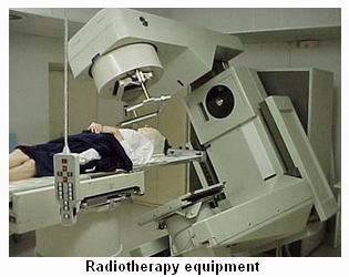
External radiotherapy is given in the radiotherapy department, using equipment similar to a large x-ray machine. Short duration radiations are given every day with rests during weekends. Treatment is individually planned for each patient, and even people with the same type of cancer may have different types of radiotherapy treatment. External radiotherapy does not make you radioactive, and it is perfectly safe for you to be with other people, including children, throughout your treatment. A course of curative (radical) treatment may be given every weekday for two to seven weeks.
Instead of having one treatment a day or having a rest at the weekend, some people will have different treatment plans. They may have more than one treatment daily, or treatment every day for two weeks. Sometimes treatment may only be given on three days each week (for example, Mondays, Wednesdays and Fridays).
There are several different types of radiotherapy machines that work in different ways. Radiotherapy treatment for most cancers, apart from skin cancers, is given by machines called linear accelerators (LinAcs).
The type of radiotherapy machine used will be carefully chosen by your specialist and physicist to give you the most appropriate treatment. Some machines are quicker than others and may give treatment in a very short time, such as a few seconds. Usually, radiotherapy treatment (including the time taken to position you) takes 10–15 minutes or less on any type of machine. The treatment itself is painless, although it may gradually cause some uncomfortable side effects.
A CT (computerized tomography) scan is first taken to access and understand the area to be treated. Multiple images from different angles are used to build up a three-dimensional picture of the area. This session will usually take about 45–60 minutes. In certain cases where a detailed study is needed an MRI would also be suggested.
Positioning
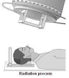
Planning for the treatment is done first, you would be positioned in a suitable couch appropriately and will have to lie very still for a few minutes so that accurate measurements can be taken and your exact position recorded. The radiographer can then make sure that you are lying in the correct position each time you have treatment. Once the treatment area has been finalized, ink markings are usually made on your skin to pinpoint the exact place where the radiation is to be directed. Sometimes three or more tattoo marks are made on the skin as they show where the radiotherapy was given and prevent further radiotherapy being given to that area in the future.
A see-through plastic mould of clear Perspex or a plastic mesh may be made; to help the body remain still when radiotherapy is given for certain parts of the body. This is often used for treatments to the head and neck area. Once you are in the correct position the staff will need to leave you alone in the room, to prevent them from being exposed to any unnecessary radiation. During treatment you will be alone for a few minutes but there will often be an intercom so that you can talk to the radiographers.
Specialized external radiotherapy techniques
Some newer ways of giving radiotherapy are being assessed to see whether they give better results than standard radiotherapy, they are:
Conformal radiotherapy uses the same radiotherapy machine as normal radiotherapy treatment. However, a device called a multi-leaf collimator which consists of a number of metal sheets which are fixed to the radiotherapy machine to arrange the beams to target the area of the cancer. Thus the stronger radiation is targeted on the tumor and the surrounding healthy tissues receive a lower dose of radiation thus reducing the side effects. Precise positioning of the radiotherapy machine is very important for conformal radiotherapy treatment and scanning is done prior to radiotherapy to ensure correct positioning.
Intensity-modulated radiotherapy (IMRT)
High-resolution intensity-modulated radiotherapy, also known as three-dimensional IMRT (3D-IMRT) also uses a multi-leaf collimator. During this treatment the layers of the multi-leaf collimator are moved while the treatment is being given. This method is able to shape the treatment beams even more precisely and allows the dose of radiotherapy to be altered in different parts of the treatment area.
Stereotactic radiotherapy
Stereotactic radiotherapy technique which directs the radiotherapy from many different angles such that the dose going to the tumor is high and the dose affecting surrounding healthy tissue is very low is used to treat brain tumors. Before treatment, several scans are analyzed by computers to ensure that the radiotherapy is precisely targeted, and the patient's head is held still in a specially-made frame while having the radiotherapy.
Stereotactic radio-surgery (gamma knife)
In fact, this type of radiotherapy, again for brain tumors, does not use a knife but very precisely targeted beams of gamma radiotherapy from hundreds of different angles. Only one session of radiotherapy, taking about four to five hours, is needed.
For this treatment you will have a specially-made metal frame attached to your head. Then several scans and x-rays are carried out to find the precise area where the treatment is needed. During the radiotherapy, you lie with your head in a large helmet, which has hundreds of holes in it to allow the radiotherapy beams through.
Internal radiotherapy is used mainly to treat cancers in the head and neck area, the cervix, womb, prostate gland or the skin.
Treatment is given by putting solid radioactive material close to or inside the tumor for a limited period of time or by using a radioactive liquid, which is given either as a drink or as an injection into a vein. In this case hospitalization is a must with special precaution taken as long as the radioactive material is in place in your body. Once the treatment is over there is no risk of exposing your family or friends to radiation.
The process of putting solid radioactive material close to or inside the tumors is called brachytherapy. Giving a radioactive liquid, either as a drink, a capsule, or as an injection into a vein is called radioisotope treatment. Before you leave hospital, the staff will check that most of the radioactivity in your body has gone, and that your belongings are free from any signs of radioactivity. After you leave hospital you should be able to carry on your life almost as normal, but there may be a few restrictions about contact with people – especially children and pregnant women.
The exact method and dosage and type of drug would be decided by your physician after necessary tests and assessments.
At the end of your treatment you will have regular follow-up appointments at the radiotherapy department or hospital. The positive effects of radiotherapy may take some time to show. People sometimes expect to be given an x-ray or a scan at the end of their treatment to see if it has worked. However, in many cases the tumour may take some time to shrink and the radiotherapy may cause some inflammation.
The other Oncology Procedures are
Few Major Hospitals for Radiotherapy are
Thailand, Malaysia, Singapore, Turkey and India are the most cost effective locations that offer up to almost 80% savings in comparison to the US.
SurgeryPlanet facilitates a plethora of services to the medical treatment traveler also which includes, a hassle free and discounted travel option, a welcome hand at the airport on arrival, travel in an air-conditioned car, round the clock service & support. Your medical evaluation is pre arranged with the least of waiting time. Once your assessment is complete and found medically fit, the procedure is immediately scheduled without a waiting period. Please read through our Services and Testimonials to understand and select your best options.
Major Treatments Abroad: Obesity / Bariatric Surgery | Spine Surgery | Stem Cell therapy | Fertility treatment | Knee replacement in India and Thailand | Heart Surgery | Organ transplant | ../ayurveda Treatment | Heart valve replacement | Hip resurfacing | Hospitals in India and Thailand for Laparoscopic Sterilization| Best hospitals in Asia | JCI & ISO certified Hospitals | Cost effective medical procedures | Healthcare tourism | Complete privacy for affordable cost | Weight loss procedures | Infertility treatment | Board certified physicians | Low cost surgeries
SurgeryPlanet is an Healthcare Facilitator and not a Medical service provider. The information provided in this website is not to be used for diagnosis or treatment of any medical condition or use for any medical purposes. We provide information solely for medical travel facilitation and do not endorse any particular health care provider, hospital, facility, destination or any healthcare service or treatment listed. We are not an agent for, or affiliated to any health care provider, or service listed in our website and is not responsible for health care services provided by them. Choice of hospital or doctor for your healthcare services is your independent decision. Consult your domestic licensed health care provider before seeking the services of any health care provider you learn about from our website.