Angiography is a medical imaging technique used to visualize the interior of blood vessels and organs. It is primarily used to detect abnormalities in the blood vessels, such as blockages, aneurysms, or vascular malformations. The procedure involves the injection of a contrast dye into the blood vessels, followed by the use of imaging technology, such as X-rays, CT (computed tomography) scans, or MRI (magnetic resonance imaging), to produce detailed images of the vascular system.
The most common type of angiography is coronary angiography, used to examine the blood vessels of the heart. However, angiography can be performed on other blood vessels throughout the body, such as those in the brain (cerebral angiography), lungs (pulmonary angiography), kidneys (renal angiography), and legs (peripheral angiography). It plays a crucial role in diagnosing a variety of vascular diseases and conditions, helping doctors determine the best course of treatment, which may include surgery, medication, or other interventions.
Angiography can be diagnostic, meaning it helps doctors identify the presence of diseases or abnormalities, or it can be therapeutic, meaning it is used to treat conditions by allowing doctors to perform procedures such as stent placement, balloon angioplasty, or clot removal while monitoring the vascular system in real-time.
While angiography is a diagnostic procedure rather than a condition, it is performed to evaluate diseases or abnormalities that affect the blood vessels. Several underlying causes and risk factors may necessitate the use of angiography:
Atherosclerosis, the buildup of plaque (a mixture of fat, cholesterol, and other substances) in the arteries, is one of the most common causes of angiography. This condition can lead to narrowing and hardening of the arteries, increasing the risk of heart disease, stroke, or peripheral artery disease (PAD). Angiography helps identify areas of blockage or restriction in the blood flow due to plaque buildup.
Coronary artery disease is caused by a buildup of plaque in the coronary arteries, which supply blood to the heart. Angiography is commonly used to assess the extent of blockages in the coronary arteries and guide treatment options such as angioplasty or bypass surgery.
Peripheral artery disease occurs when there is a narrowing or blockage of the arteries supplying blood to the legs and feet. Angiography can help identify blockages in these arteries, allowing doctors to determine whether surgical intervention or stent placement is necessary.
An aneurysm is an abnormal bulge in a blood vessel caused by a weakening of the vessel wall. Aneurysms can occur in any blood vessel but are most common in the aorta, brain, and legs. Angiography is used to detect aneurysms and assess their size and location, which helps doctors determine the appropriate treatment, including surgery.
A stroke can occur due to a blockage or rupture of blood vessels in the brain. Angiography, specifically cerebral angiography, is used to evaluate blood flow in the brain, detect blockages, and identify abnormal vessels that may require treatment.
Pulmonary embolism occurs when a blood clot obstructs a pulmonary artery, cutting off blood flow to the lungs. Pulmonary angiography is the gold standard for diagnosing pulmonary embolism, as it provides detailed imaging of the arteries in the lungs.
Certain individuals may be born with vascular malformations, where the blood vessels form abnormally. Angiography is used to identify these malformations and help guide treatment or intervention.
Various factors increase the risk of developing conditions that may require angiography, including:
High blood pressure (hypertension)
High cholesterol levels
Diabetes
Obesity
Smoking
Family history of heart disease or stroke
Sedentary lifestyle
Angiography is not a condition itself but a diagnostic procedure performed when there are signs or symptoms suggesting problems with the blood vessels. Common symptoms and signs that may lead to the need for angiography include:
Chest pain (angina) or discomfort in the chest is a common symptom of coronary artery disease (CAD) and may indicate a blockage in the coronary arteries. Angiography is used to evaluate the severity of the blockages and determine whether interventions like angioplasty or stent placement are necessary.
Shortness of breath or difficulty breathing may be a sign of heart disease or pulmonary embolism. If this symptom is present along with chest pain, swelling, or leg pain, angiography can help assess whether blood clots, blockages, or other vascular issues are affecting the heart or lungs.
Pain, cramping, or fatigue in the legs, particularly when walking, can be a sign of peripheral artery disease (PAD), where the arteries supplying blood to the legs become narrowed or blocked. Angiography is used to evaluate the arteries in the legs and determine if balloon angioplasty or bypass surgery is needed.
Sudden weakness, numbness, or paralysis on one side of the body could be a sign of a stroke or cerebrovascular disease. Angiography can help identify areas of the brain that are lacking adequate blood supply due to blocked or narrowed arteries.
A severe headache, blurred vision, or difficulty speaking can be symptoms of a cerebral aneurysm or vascular malformation in the brain. Angiography can help detect these issues and guide appropriate treatment to prevent further damage.
Swelling, pain, or redness in the legs may indicate deep vein thrombosis (DVT) or pulmonary embolism. Angiography can help identify the presence of blood clots in the veins or arteries and assist in determining the appropriate treatment.
Angiography is typically recommended when non-invasive imaging techniques (such as ultrasound, CT scans, or MRI) are insufficient or inconclusive. The diagnostic process usually involves several steps:
The healthcare provider will begin by reviewing the patient's medical history, including any symptoms, risk factors (e.g., smoking, high blood pressure, or diabetes), and family history of vascular disease. The doctor will also perform a physical examination to assess the patient's overall health and signs of cardiovascular or vascular conditions.
Before proceeding with angiography, the doctor may recommend non-invasive imaging tests, such as:
Ultrasound: A non-invasive test used to visualize blood flow in the arteries and veins.
CT Scan or MRI: Both can provide detailed images of the blood vessels and surrounding structures, though they may not offer the same level of detail as angiography in some cases.
Once the decision is made to proceed with angiography, the procedure typically follows these steps:
Contrast Injection: A special dye (contrast agent) is injected into the blood vessels through a catheter, usually inserted in the groin or arm.
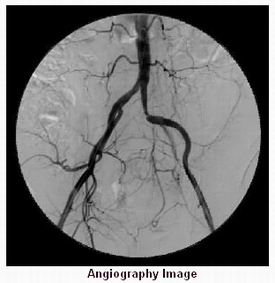
Imaging: As the contrast dye flows through the blood vessels, the physician uses imaging technology such as X-rays, CT scans, or MRI to capture images of the vascular system. These images can reveal blockages, narrowing, aneurysms, or other abnormalities.
Once the images are obtained, a radiologist or cardiologist will analyze the results. The findings will help guide the diagnosis and treatment plan, whether it involves surgery, medications, or lifestyle modifications.
While angiography itself is a diagnostic tool, it is often used in conjunction with treatment options aimed at managing vascular conditions. Treatment options depend on the findings from the angiography and the underlying condition:
Balloon angioplasty involves the use of a catheter with a balloon at its tip, which is inflated at the site of the blockage to widen the artery. This procedure is commonly used to treat coronary artery disease and peripheral artery disease.
After angioplasty, a stent (a small mesh tube) may be placed in the artery to help keep it open. Stenting is commonly used in coronary artery disease and peripheral artery disease to prevent the artery from collapsing or narrowing again.
In more severe cases, surgery may be necessary to remove blockages or bypass narrowed arteries. Coronary artery bypass grafting (CABG) is a common procedure for patients with severe coronary artery disease, while bypass surgery can also be performed for peripheral artery disease.
Medications such as antiplatelet drugs (aspirin or clopidogrel) or blood thinners (warfarin) are commonly prescribed to reduce the risk of blood clot formation, especially after angioplasty or stent placement. Statins may also be prescribed to lower cholesterol levels and reduce the risk of atherosclerosis.
Preventing the need for angiography involves addressing the underlying risk factors for cardiovascular and vascular diseases. Management strategies include:
Diet: Eating a healthy, balanced diet rich in fruits, vegetables, whole grains, and lean proteins can reduce the risk of heart disease, stroke, and peripheral artery disease.
Exercise: Regular physical activity, such as walking, swimming, or cycling, can help maintain a healthy weight, reduce blood pressure, and improve circulation.
Smoking Cessation: Quitting smoking is one of the most important steps in reducing the risk of vascular diseases.
Managing underlying conditions such as hypertension, diabetes, and high cholesterol can help reduce the risk of developing vascular diseases. Regular monitoring of these conditions can prevent complications and reduce the need for invasive procedures like angiography.
Angiography is generally a safe procedure, but as with any medical intervention, there are potential risks and complications, including:
Bleeding at the catheter insertion site is one of the most common complications of angiography. Careful monitoring and pressure on the site after the procedure can reduce the risk of excessive bleeding.
As with any invasive procedure, there is a risk of infection at the catheter insertion site. Antibiotics may be administered to prevent infection.
Some individuals may have allergic reactions to the contrast dye used during angiography. Symptoms can include rash, itching, or difficulty breathing. Pre-procedure screening can help identify those at risk.
The contrast dye used in angiography can sometimes affect kidney function, particularly in individuals with pre-existing kidney disease. Hydration before and after the procedure can help minimize this risk.
After angiography, the lifestyle and management of the underlying condition will depend on the results of the procedure. Patients may need to:
Regular follow-up appointments with healthcare providers will help monitor any changes in the vascular system. If interventions like angioplasty or stent placement were performed, ongoing monitoring will ensure the success of the treatment.
Adhering to lifestyle modifications such as maintaining a healthy diet, exercising regularly, and managing stress is essential to prevent further vascular problems and reduce the risk of complications.
Dealing with conditions requiring angiography, such as heart disease or stroke, can be emotionally challenging. Support groups, therapy, and counseling can help individuals manage stress and anxiety related to their diagnosis and treatment.
Angiography is a medical imaging technique used to visualize the inside of blood vessels and organs, such as the heart, brain, and kidneys, to check for any abnormalities or blockages. It involves the injection of a contrast dye (also known as a contrast agent) into the blood vessels, which helps make the vessels visible on X-ray or other imaging methods like CT (computed tomography) or MRI (magnetic resonance imaging). Angiography is commonly used to diagnose conditions like coronary artery disease, aneurysms, and vascular blockages.
Angiography is performed to:
Identify blockages: Detect narrowing, blockages, or other abnormalities in the blood vessels, often in the coronary arteries of the heart or the arteries supplying the brain and kidneys.
Evaluate aneurysms: Identify any abnormal dilations or bulges in the blood vessels.
Diagnose vascular diseases: Such as peripheral artery disease (PAD) or atherosclerosis (hardening of the arteries).
Assess organ blood flow: To examine how well blood is flowing to specific organs, such as the brain, heart, or kidneys.
Guide treatment: It can also be used to guide certain treatments like stent placement, balloon angioplasty, or the removal of a clot.
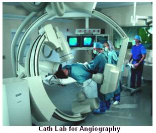
Angiography is typically performed using a catheter inserted through the skin, usually in the groin or wrist. The general process involves:
Preparation: The patient is given a mild sedative to help them relax, and the area where the catheter will be inserted is cleaned and numbed.
Insertion of catheter: A small tube (catheter) is inserted into an artery, often in the groin or wrist, and guided to the area being examined.
Injection of contrast dye: A contrast dye is injected through the catheter into the blood vessels. This makes the vessels visible on X-ray or other imaging techniques.
Imaging: X-rays or other imaging scans are taken to capture images of the blood vessels. The doctor can analyze the images to assess blood flow, blockages, or abnormalities.
Post-procedure: The catheter is removed, and the insertion site is bandaged. You may be asked to lie still for several hours afterward to prevent bleeding.
The procedure typically takes 30 minutes to 1 hour depending on the area being examined.
Angiography is generally not painful. The patient may experience some discomfort when the catheter is inserted, as the doctor will need to numb the area. The injection of the contrast dye may cause a warm sensation or a brief metallic taste in the mouth, but this is typically short-lived. After the procedure, some mild soreness or bruising may occur at the catheter insertion site, but it is usually manageable with pain relief if necessary.
While angiography is generally safe, there are some potential risks and complications, including:
Allergic reaction: A mild or severe allergic reaction to the contrast dye used in the procedure.
Bleeding or hematoma: At the catheter insertion site, there is a risk of bleeding or the formation of a bruise (hematoma).
Infection: Though rare, there is a risk of infection at the catheter insertion site.
Damage to blood vessels: The catheter could accidentally cause injury to blood vessels, leading to further complications.
Kidney damage: In some cases, the contrast dye can cause kidney damage, especially in patients with pre-existing kidney problems.
Blood clots: There is a risk of blood clot formation, which can travel to other parts of the body and cause problems like stroke or heart attack.
Your doctor will assess the risks based on your health history and take precautions to minimize complications.
Before undergoing angiography, you will need to follow certain preparation guidelines:
Fasting: You may be asked to avoid eating or drinking for 6-8 hours before the procedure.
Medications: Inform your doctor about any medications you are taking, including blood thinners or diabetes medications. You may need to adjust or temporarily stop certain medications before the procedure.
Allergy check: Be sure to inform your doctor if you have any allergies, especially to iodine or contrast dyes, as this could affect the procedure.
Inform the medical team: Let the healthcare team know if you have any kidney problems, diabetes, or heart conditions.
Angiography usually takes 30 minutes to 1 hour. The actual procedure may be shorter or longer depending on the complexity of the examination and whether additional treatments (such as stent placement) are performed during the procedure. You may also need to spend some time in recovery afterward to monitor your condition and ensure there are no complications.
After the procedure, you will be monitored for a short time, typically 1 to 2 hours, to ensure that there are no immediate complications, such as bleeding or allergic reactions. The catheter insertion site will be bandaged, and you may be asked to lie still for several hours to allow the blood vessel to heal. Your doctor will provide instructions on caring for the site and any restrictions on activity.
Most patients can go home the same day, but you may be advised to avoid strenuous activity for a few days. You should follow your doctor’s post-procedure care instructions closely.
The results of angiography are usually available immediately after the procedure. The imaging is reviewed by a radiologist or a specialist, and the findings are sent to your doctor. Depending on the purpose of the angiography, your doctor will discuss the results with you and determine the next steps for diagnosis or treatment.
Yes, angiography is not only a diagnostic tool but also a therapeutic procedure in some cases. If the angiogram reveals blockages or other issues, treatments can be performed during the same procedure, such as:
Stent placement: A stent can be inserted to open up narrowed or blocked blood vessels.
Balloon angioplasty: A balloon is inflated inside a narrowed blood vessel to widen it and restore normal blood flow.
Thrombolysis: In some cases, blood clots can be treated with medications delivered through the catheter.
This ability to treat the problem immediately during the angiogram is one of the advantages of this procedure.
The other Radiology Procedures are:
Few Major Hospitals for Angiography are:
Thailand, Malaysia, Singapore, Turkey and India are the most cost effective locations that offer up to almost 80% savings in comparison to the US.
SurgeryPlanet facilitates a plethora of services to the medical treatment traveler also which includes, a hassle free and discounted travel option, a welcome hand at the airport on arrival, travel in an air-conditioned car, round the clock service & support. Your medical evaluation is pre arranged with the least of waiting time. Once your assessment is complete and found medically fit, the procedure is immediately scheduled without a waiting period. Please read through our Services and Testimonials to understand and select your best options.
Major Treatments Abroad: Obesity / Bariatric Surgery | Spine Surgery | Stem Cell therapy | Fertility treatment | Knee replacement in India and Thailand | Heart Surgery | Organ transplant | Ayurveda Treatment | Heart valve replacement | Hip resurfacing | Hospitals in India and Thailand for Laparoscopic Sterilization| Best hospitals in Asia | JCI & ISO certified Hospitals | Cost effective medical procedures | Healthcare tourism | Complete privacy for affordable cost | Weight loss procedures | Infertility treatment | Board certified physicians | Low cost surgeries
SurgeryPlanet is an Healthcare Facilitator and not a Medical service provider. The information provided in this website is not to be used for diagnosis or treatment of any medical condition or use for any medical purposes. We provide information solely for medical travel facilitation and do not endorse any particular health care provider, hospital, facility, destination or any healthcare service or treatment listed. We are not an agent for, or affiliated to any health care provider, or service listed in our website and is not responsible for health care services provided by them. Choice of hospital or doctor for your healthcare services is your independent decision. Consult your domestic licensed health care provider before seeking the services of any health care provider you learn about from our website.