Microsurgical discectomy is a highly effective surgical procedure aimed at treating patients suffering from herniated discs, commonly known as slipped discs, which lead to significant nerve compression. This surgery, performed using advanced microsurgical techniques, allows spinal surgeons to remove the damaged portion of a disc that presses against the nerves or spinal cord, thereby providing relief from pain, numbness, and weakness caused by the compression.
Microsurgical discectomy is a preferred option for patients experiencing severe symptoms that do not improve with conservative treatments like physical therapy, medications, or epidural injections. The microsurgical technique involves minimally invasive methods, which reduce tissue disruption and lead to faster recovery times compared to traditional open surgery.
This guide aims to provide a comprehensive understanding of microsurgical discectomy, its causes, treatment options, post-operative care, and how to manage the condition effectively.
A herniated disc occurs when the inner, jelly-like core of the spinal disc (nucleus pulposus) pushes through a tear or rupture in the outer layer (annulus fibrosus). The herniated portion of the disc can then compress surrounding nerves, causing pain and functional impairments. Some common causes of a herniated disc include:
Age-Related Degeneration (Degenerative Disc Disease):
As we age, the spinal discs lose hydration and elasticity, making them more prone to damage and herniation. This is the most common cause of herniated discs in older adults.
Trauma or Injury:
Sudden impacts or injuries from car accidents, sports, or falls can cause discs to rupture, leading to nerve compression and herniation.
Repetitive Strain or Overuse:
Occupations or activities that involve frequent heavy lifting, twisting, or prolonged sitting may increase the risk of disc herniation over time.
Genetics:
Some individuals inherit weaker disc structures or a predisposition to disc herniation, making them more vulnerable to developing this condition.
Poor Posture and Body Mechanics:
Poor posture and incorrect lifting techniques can place extra stress on the spine and increase the likelihood of a herniated disc.
Age: Most commonly affects individuals between the ages of 30 and 50.
Occupational Hazards: Jobs that involve heavy lifting, bending, or twisting.
Obesity: Excess weight puts more pressure on the spine and increases the risk.
Smoking: Reduces oxygen supply to the spine, impairing the healing of discs.
Sedentary Lifestyle: Lack of physical activity can weaken the muscles supporting the spine.
The primary symptoms of a herniated disc are the result of nerve compression. These symptoms can vary depending on the location of the herniation, but common signs include:
Severe Back or Neck Pain: Pain that radiates from the back or neck to the arms or legs, often described as a deep ache or burning sensation.
Muscle Weakness: Weakness in muscles controlled by the compressed nerve, which can lead to difficulty walking or moving limbs.
Numbness or Tingling: A sensation of "pins and needles" in the arms, hands, legs, or feet.
Sciatica: Radiating pain from the lower back to the buttocks, thighs, and legs, often on one side.
Cervical Radiculopathy: Pain radiating from the neck down to the arms and hands.
Loss of Reflexes: Decreased or absent reflexes in the affected area.
Bladder or Bowel Dysfunction: In rare and severe cases, compression can lead to difficulty controlling bladder or bowel movements.
If these symptoms persist or worsen, microsurgical discectomy is often considered to relieve pressure on the nerves and provide long-term relief.
The diagnosis of a herniated disc requiring microsurgical discectomy involves a combination of clinical evaluation and imaging techniques.
Patient History: A detailed review of symptoms, their onset, and any contributing activities or injuries.
Physical and Neurological Examination: The doctor will assess muscle strength, reflexes, and sensation in the arms or legs, checking for signs of nerve compression.
MRI (Magnetic Resonance Imaging): The gold standard for diagnosing disc herniation, providing detailed images of the discs, spinal cord, and nerves.
CT Scan (with Myelogram): Offers detailed images of the spine and helps identify disc herniation in patients who may not be candidates for MRI.
X-rays: Although they do not show soft tissues, they can be used to rule out other conditions like fractures or misalignments.
Electromyography (EMG): Measures electrical activity in muscles to assess nerve function and detect nerve compression.
Accurate diagnosis is essential to ensure that microsurgical discectomy is the most appropriate treatment option.
When conservative treatments fail, microsurgical discectomy is an effective surgical option for patients suffering from a herniated disc. The goal of the surgery is to remove the part of the disc that is pressing on the nerve, thereby relieving pain and improving function.
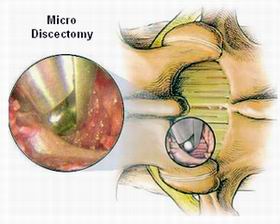
Minimally Invasive Technique: Microsurgical discectomy uses small incisions (typically 1–2 cm) and specialized instruments, including a microscope for enhanced visualization of the disc and nerves.
Removal of Herniated Disc: The damaged portion of the disc is carefully removed, decompressing the nerve while preserving as much of the disc as possible.
Minimized Tissue Disruption: Unlike traditional open surgery, the minimally invasive nature of microsurgical discectomy reduces damage to surrounding tissues, promoting faster recovery and fewer complications.
Non-surgical Treatments (Initial approach):
Physical Therapy: Focused on strengthening muscles and improving spinal flexibility.
Pain Management: Use of non-steroidal anti-inflammatory drugs (NSAIDs) or stronger pain relievers.
Epidural Steroid Injections: To reduce inflammation and alleviate pain.
Other Surgical Approaches:
Laminectomy: Removal of part or all of the lamina (bony arch of the vertebra).
Spinal Fusion: Fusing two vertebrae together to prevent movement at the site of the disc removal.
While not all cases of herniated discs can be prevented, there are preventive measures and management strategies that can reduce the risk or promote better recovery after surgery.
Proper Posture: Maintaining a neutral spine while sitting, standing, and sleeping.
Correct Lifting Techniques: Bending at the knees, not the back, when lifting heavy objects.
Regular Exercise: Strengthening the core and back muscles can help support the spine.
Healthy Weight: Reducing excess body weight reduces pressure on the spine.
Avoid Smoking: Smoking accelerates disc degeneration by reducing blood flow to the spine.
Physical Therapy: Tailored rehabilitation to improve spinal stability and mobility.
Pain Management: Use of prescribed medications during the recovery phase.
Posture Correction: Maintaining good posture to reduce strain on the spine.
Long-term Monitoring: Periodic follow-ups with the surgeon to ensure proper healing and prevent reherniation.
Although microsurgical discectomy is considered a safe procedure, there are some potential complications that patients should be aware of:
Nerve Injury: Although rare, there is a risk of nerve damage during the removal of the herniated disc.
Infection: Any surgical procedure carries a small risk of infection at the incision site.
Recurrent Disc Herniation: The disc may herniate again at the same or adjacent level.
Cerebrospinal Fluid (CSF) Leak: Rarely, a tear in the dura (the covering of the spinal cord) can occur during surgery.
Blood Clots: Patients may be at risk for developing blood clots post-surgery due to immobility.
Most complications are rare and can be minimized with proper surgical techniques and post-operative care.
After undergoing microsurgical discectomy, patients can expect a significant reduction in pain and a faster recovery compared to traditional surgery. However, the road to full recovery can take several months.
First Few Weeks: Limited activity and pain management are essential.
1–3 Months: Gradual return to normal activities, with physical therapy and rehabilitation.
6–12 Months: Full recovery, with ongoing exercises to maintain a healthy back.
Patients are advised to maintain a healthy lifestyle and engage in preventive measures to avoid future back problems. Avoiding heavy lifting, staying active, and practicing proper posture are all essential components of post-surgical life.
Dealing with chronic pain before surgery can be mentally and emotionally taxing.
Post-surgery recovery can also bring challenges, but understanding the recovery process and receiving emotional support can help patients regain confidence and return to normal life.
Microsurgical discectomy is a minimally invasive surgical procedure used to treat herniated or slipped discs in the spine, typically in the lower back (lumbar spine) or neck (cervical spine). It involves the removal of the part of the disc that is pressing on the nerves, causing pain or other neurological symptoms. The procedure is performed using a microscope, allowing the surgeon to view the area with high precision while making small incisions.
Microsurgical discectomy uses advanced techniques, including a microscope and smaller incisions, to remove the herniated disc material. This results in less muscle disruption, reduced blood loss, and a quicker recovery time compared to traditional discectomy, which requires larger incisions and greater disruption of surrounding tissues.
Candidates for microsurgical discectomy typically suffer from a herniated disc that is compressing a nerve and causing symptoms such as back pain, leg pain (sciatica), numbness, or weakness. The procedure is considered when non-surgical treatments, such as physical therapy or medications, have failed. Ideal candidates are those in good overall health with a single, localized disc herniation.
The benefits of microsurgical discectomy include:
Smaller incisions and minimal tissue damage
Faster recovery time compared to traditional surgery
Lower risk of complications like infection or blood loss
Quicker return to normal activities and work
Preservation of spine stability with no need for fusion in most cases
While microsurgical discectomy is generally safe, potential risks include:
Infection at the incision site
Nerve damage
Persistent pain or recurrence of herniated disc
Dural tear or cerebrospinal fluid leakage
Blood clots
However, these risks are rare, and the procedure is associated with a high success
rate.
Recovery from microsurgical discectomy is typically faster than traditional surgery. Most patients can return to light activities within 1-2 weeks. Full recovery, including the resumption of heavy lifting or strenuous exercise, may take 6 to 8 weeks. Physical therapy is often recommended to help strengthen the muscles around the spine and promote healing.
Some discomfort or mild pain may be experienced after the surgery, especially during the initial recovery period. However, most patients report significant relief from their pre-surgery symptoms, such as leg pain or numbness. Pain management strategies, including medications and physical therapy, are used to control any post-operative discomfort.
Yes, microsurgical discectomy can be performed on both the cervical spine (neck) and the lumbar spine (lower back). The procedure is performed based on the location of the herniated disc, and the approach may vary slightly depending on the region being treated.
Microsurgical discectomy typically takes between 1 to 2 hours, depending on the complexity of the herniated disc and the specific area being treated. The procedure is minimally invasive, so it can often be completed relatively quickly compared to traditional discectomy surgeries.
Most patients stay in the hospital for 1 to 2 days after microsurgical discectomy. During this time, pain is managed with medications, and you will be encouraged to begin walking as soon as possible. The medical team will monitor you for any complications and ensure you are comfortable before discharge. You will receive instructions on caring for the incision site, activity restrictions, and follow-up appointments.

The other Neurology Procedures are:
Few Major Hospitals for Microsurgical Discectomy are
Thailand, Malaysia, Singapore, Turkey and India are the most cost effective locations that offer up to almost 80% savings in comparison to the US.
SurgeryPlanet facilitates a plethora of services to the medical treatment traveler also which includes, a hassle free and discounted travel option, a welcome hand at the airport on arrival, travel in an air-conditioned car, round the clock service & support. Your medical evaluation is pre arranged with the least of waiting time. Once your assessment is complete and found medically fit, the procedure is immediately scheduled without a waiting period. Please read through our Services and Testimonials to understand and select your best options.
Major Treatments Abroad: Obesity / Bariatric Surgery | Spine Surgery | Stem Cell therapy | Fertility treatment | Knee replacement in India and Thailand | Heart Surgery | Organ transplant | Ayurveda Treatment | Heart valve replacement | Hip resurfacing | Hospitals in India and Thailand for Laparoscopic Sterilization| Best hospitals in Asia | JCI & ISO certified Hospitals | Cost effective medical procedures | Healthcare tourism | Complete privacy for affordable cost | Weight loss procedures | Infertility treatment | Board certified physicians | Low cost surgeries
SurgeryPlanet is an Healthcare Facilitator and not a Medical service provider. The information provided in this website is not to be used for diagnosis or treatment of any medical condition or use for any medical purposes. We provide information solely for medical travel facilitation and do not endorse any particular health care provider, hospital, facility, destination or any healthcare service or treatment listed. We are not an agent for, or affiliated to any health care provider, or service listed in our website and is not responsible for health care services provided by them. Choice of hospital or doctor for your healthcare services is your independent decision. Consult your domestic licensed health care provider before seeking the services of any health care provider you learn about from our website.